
Advanced Imaging
 Advances in digital radiology allow us to x-ray our patients safely and quickly. Because a digital x-ray system requires no film, it reduces patient radiation exposure and eliminates hazardous waste. Patients simply close their mouths on digital sensors—instead of on film—while computers process the imagery. A quick, outside of the mouth scan is all it takes. Patients will not have to suffer through 18-22 uncomfortable individual inside-the-mouth x-rays. This is especially great news for people with a high gag reflex, small mouths or who have difficulty taking conventional x-rays. Dr. Myers and his trained staff know the correct positioning to take a single exposure (which uses less radiation than a full mouth series of x-rays) producing the best quality image. Within seconds, the digital images are ready for viewing.
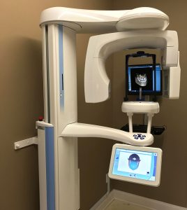
Doctors striving for excellence in patient care know that the more information they have about the anatomy of their patient’s condition the better. That’s why Dr. Myers uses Planmeca as a cone beam x-ray that brings in the 3rd dimension. He is one of the few general dentists in the region to be using Cone-Beam 3D imaging. 3D imaging provides a diagnostic, extremely low-dose x-ray for use in a wide variety of procedures. Nearly all implant dentistry requires 3D imaging. Most practices need to refer patients out of office for costly higher radiation images provided by a medical imaging center. Dr. Myers is able to provide the service at a very low cost. The 3D scan is important for planning implant surgeries as well as diagnosing and determining findings that cannot be seen on routine x-rays. It provides a visual reconstruction of the patient’s anatomy that is comparable to traditional medical CT scans, but costs less and uses significantly less radiation – only slightly more radiation that a Panorex film. A single exposure provides a basic panoramic view, a lateral skull view, and literally hundreds of other views. The images can be magnified, rotated in space, viewed from any angle, sliced and sectioned to improve visualization in 3D. Dr. Myers is no longer limited to making an “educated guess” off of a 2-D x-ray. With the Planmeca images, Dr. Myers can see inside the bone and teeth in 3-D, making diagnosis more exact and accurate. This translates more predictable and safer treatments outcomes. In addition, the ability to scan through teeth and bone in 3-D allows for more accurate diagnosis of infection, root fractures, abscesses cysts and tumors or any problems that were difficult or impossible to diagnose with a 2-D x-ray.
If you have lingering pain or a problem that eluded diagnosis, we welcome you to take advantage of superior diagnosis with our advanced imaging technology.